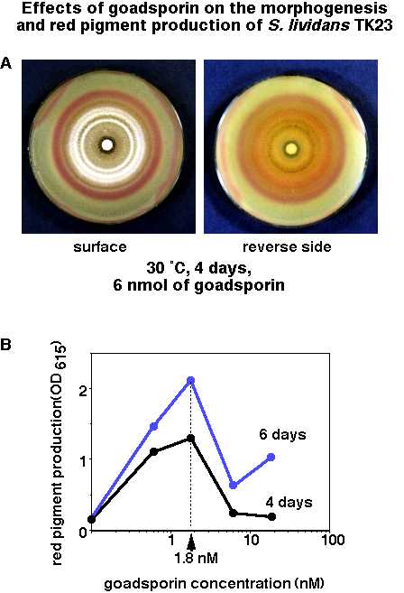
Fig. 2. Effects of goadsporin on the morphogenesis and red pigment
production of S. lividans TK23 on solid medium (A) and effects of
goadsporin on the red pigment production by S. lividans TK23 on
liquid medium (B).
(A) Photographs were taken after 4 days of growth on Bennett's
agar medium. 6 nmol (ca. 10 micro g) of goadsporin was absorbed in paperdiscs.
Left: a photograph of the surface of the plate. Aerial mycerium formation
was observed in the white ring zone, and Spore formation was observed in
the gray zone which located inside of the white zone. Right: a photograph
of the reverse side of the plate. Red pigment production was observed
around the paper disc. (B) Actinorhodin production in Bennett's liquid
medium containing various concentration of goadsporin. Production
after 4 days growth (black) and 6 days growth (grey) was determined by
following A615 of the culture broth at pH 12.